Pubblicazioni

Cerebral atrophy and white matter disruption in chronic schizophrenia  (2007)

Autori:
Andreone, N; Tansella, Michele; Cerini, Roberto; Rambaldelli, Gianluca; Versace, Amelia; Marrella, Giovanna; Perlini, Cinzia; Dusi, Nicola; Pelizza, L; Balestrieri, M; Barbui, Corrado; Nose', Michela; Gasparini, A; Brambilla, P.
Titolo:
Cerebral atrophy and white matter disruption in chronic schizophrenia
Anno:
2007
Tipologia prodotto:
Articolo in Rivista
Tipologia ANVUR:
Articolo su rivista
Lingua:
Inglese
Formato:
A Stampa
Referee:
Nome rivista:
European Archives of Psychiatry and Clinical Neuroscience
ISSN Rivista:
0940-1334
N° Volume:
257
Numero o Fascicolo:
1
Intervallo pagine:
3-11
Parole chiave:
magnetic resonance; diffusion imaging; neuroimaging; brain imaging; ventricles; cerebellum; lobes
Breve descrizione dei contenuti:
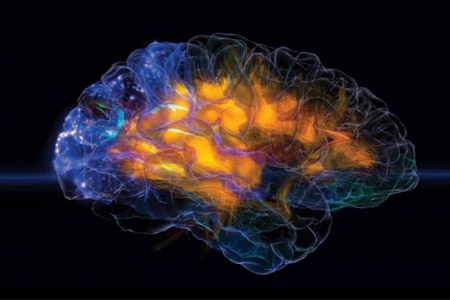
Several magnetic resonance imaging (MRI) studies have shown cerebral atrophy in established schizophrenia, although not in all reports. Discrepancies may mostly be due to population and postprocessing differences. Recently, disruption of cortical white matter integrity has also been reported in chronic patients with schizophrenia. In this study we explored tridimensional (3D) cerebral volumes and white matter microstructure in schizophrenia with structural and diffusion magnetic resonance. Twenty-five patients with established schizophrenia and 25 1:1 matched normal controls underwent a session of MRI using a Siemens 1.5T-scanner. 3D brain volume reconstruction was performed with the semi-automatic software Amira (TGS, San Diego, CA), whereas the apparent diffusion coefficients (ADCs) of cortical white matter water molecules were obtained with in-house developed softwares written in MatLab (The Mathworks-Inc., Natick, MA). Compared to controls, patients with schizophrenia had significantly smaller gray matter intracranium and total brain volumes, increased 4th ventricle volumes, and greater temporal and occipital ADCs. Patients treated with typical antipsychotic medication (N = 9) had significantly larger right lateral and 4th ventricles compared to those on atypical antipsychotic drugs. Intracranial volumes significantly inversely correlated with left temporal ADC in patients with schizophrenia. Also, age correlated directly with right, left, and 3rd ventricle volumes and inversely with gray matter intracranium volumes in individuals with schizophrenia. This study confirmed the presence of cortical atrophy in patients with schizophrenia, especially in those on typical antipsychotic drugs, and the existence of white matter disruption. It also suggested that physiological aging effects on brain anatomy may be abnormally pronounced in schizophrenia.
Id prodotto:
43733
Handle IRIS:
11562/308831
depositato il:
5 dicembre 2007
ultima modifica:
15 novembre 2022
Citazione bibliografica:
Andreone, N; Tansella, Michele; Cerini, Roberto; Rambaldelli, Gianluca; Versace, Amelia; Marrella, Giovanna; Perlini, Cinzia; Dusi, Nicola; Pelizza, L; Balestrieri, M; Barbui, Corrado; Nose', Michela; Gasparini, A; Brambilla, P., Cerebral atrophy and white matter disruption in chronic schizophrenia «European Archives of Psychiatry and Clinical Neuroscience» , vol. 257 , n. 12007pp. 3-11

Consulta la scheda completa presente nel repository istituzionale della Ricerca di Ateneo IRIS

<<indietro

Attività

Strutture

Condividi