- Autori:
-
M. S. Buchsbaum;M. Haznedar;R. E. Newmark;K. Chu;N. Dusi;J. J. Entis;K. E. Goldstein;C. R. Goodman;A. Gupta;E. Hazlett;J. Iannuzzi;Y. Torosjan;J. Zhang;A. Wolkin
- Titolo:
-
FDG-PET and MRI imaging of the effects of sertindole and haloperidol in the prefrontal lobe in schizophrenia.
- Anno:
-
2009
- Tipologia prodotto:
-
Articolo in Rivista
- Tipologia ANVUR:
- Articolo su rivista
- Lingua:
-
Inglese
- Formato:
-
A Stampa
- Referee:
-
Sì
- Nome rivista:
- Schizophrenia Research
- ISSN Rivista:
- 0920-9964
- N° Volume:
-
114
- Intervallo pagine:
-
161-171
- Codice PMID:
-
19695836
- Parole chiave:
-
Adult, Analysis of Variance, Antipsychotic Agents, pharmacology/therapeutic use, Brain Mapping, Female, Fluorodeoxyglucose F18, diagnostic use, Frontal Lobe, drug effects/pathology/radionuclide imaging, Haloperidol, Humans, Imidazoles, Indoles, Magnetic Resonance Imaging, methods, Male, Middle Aged, Neuropsychological Tests, Positron-Emission Tomography, Psychiatric Status Rating Scales, Schizophrenia, drug therapy/pathology/radionuclide imaging, Young Adult
- Breve descrizione dei contenuti:
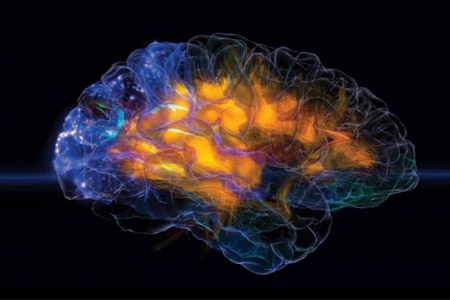
- Sertindole, a 2nd generation antipsychotic with low movement disorder side effects, was compared with haloperidol in a 6-week crossover study. Fifteen patients with schizophrenia (mean age=42.6, range=22-59, 11 men and 4 women) received sertindole (12-24 mg) or haloperidol (4-16 mg) for 6 weeks and then received a FDG-PET scan and an anatomical MRI. Patients were then crossed to the other treatment and received a second set of scans at week 12. Dose was adjusted by a physician blind to the medication type. Brodmann areas were identified stereotaxically using individual MRI templates applied to the coregistered FDG-PET image. Sertindole administration was associated with higher dorsolateral prefrontal cortex metabolic rates than haloperidol and lower orbitofrontal metabolic rates than haloperidol. This effect was greatest for gray matter of the dorsolateral Brodmann areas 8, 9, 10, 44, 45, and 46. Patients were further contrasted with an approximately age and sex-matched group of 33 unmedicated patients with schizophrenia and with a group of 55 normal volunteers. Sertindole administration was associated with greater change toward normal values and away from the values found in the unmedicated comparison group for dorsolateral prefrontal cortex gray matter and white matter underlying medial prefrontal and cingulate cortex. These results are consistent with the low motor side-effect profile of sertindole, greater improvement on prefrontal cognitive tasks with sertindole than haloperidol, and with the tendency of 2nd generation antipsychotic drugs to have greater frontal activation than haloperidol.
- Pagina Web:
-
http://dx.doi.org/10.1016/j.schres.2009.07.015
- Id prodotto:
-
61554
- Handle IRIS:
-
11562/361610
- depositato il:
-
26 agosto 2011
- ultima modifica:
-
2 novembre 2016
- Citazione bibliografica:
-
M. S. Buchsbaum;M. Haznedar;R. E. Newmark;K. Chu;N. Dusi;J. J. Entis;K. E. Goldstein;C. R. Goodman;A. Gupta;E. Hazlett;J. Iannuzzi;Y. Torosjan;J. Zhang;A. Wolkin,
FDG-PET and MRI imaging of the effects of sertindole and haloperidol in the prefrontal lobe in schizophrenia.
«Schizophrenia Research»
, vol.
114
,
2009
,
pp. 161-171
Consulta la scheda completa presente nel
repository istituzionale della Ricerca di Ateneo